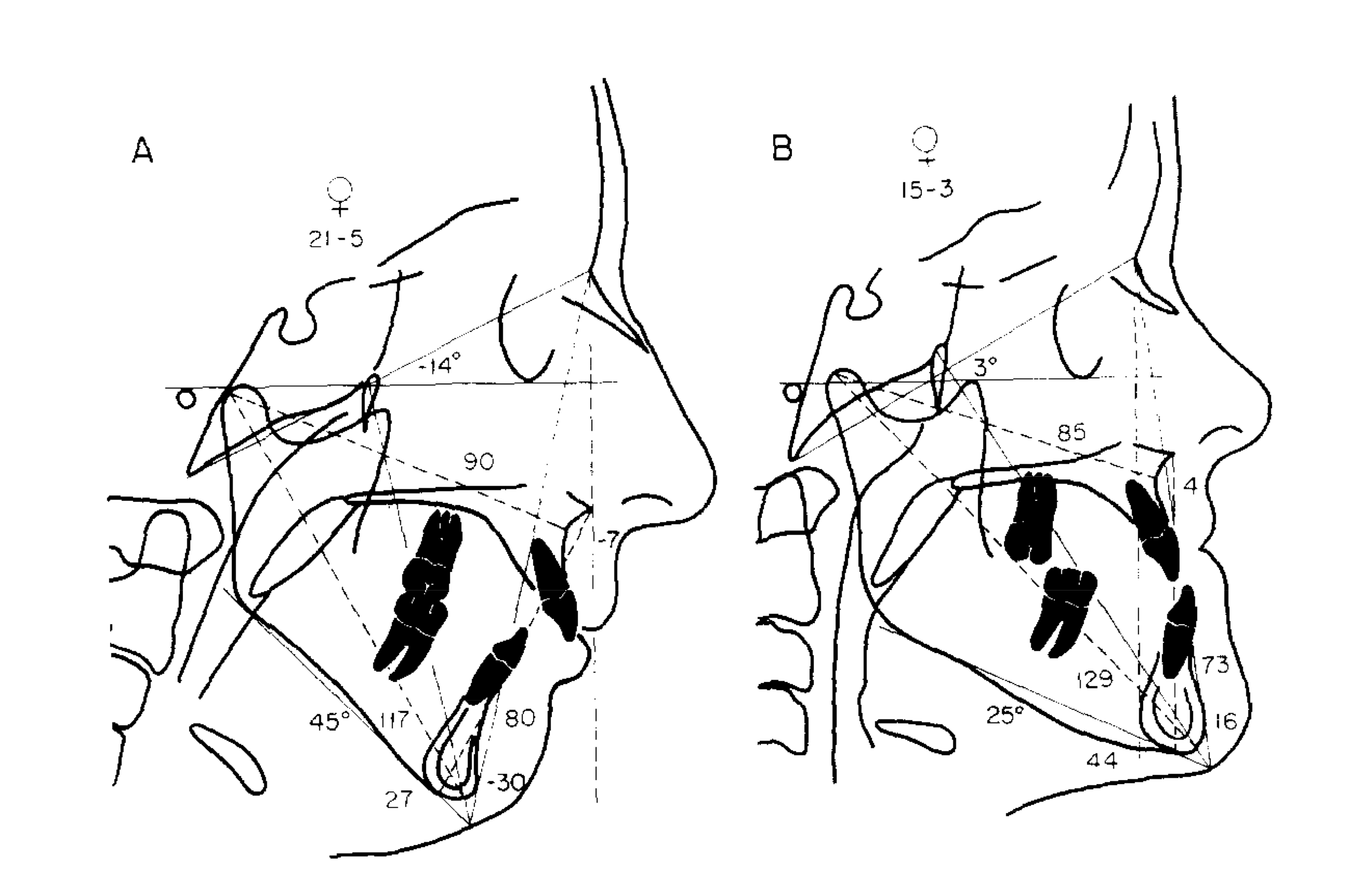
The analysis is composed of preset measurements of angles and lengths based on standard values of three cephalometric sample groups. The first sample was derived from lateral cephalograms of young patients with Bolton standards. The second sample consists of values from a group of normal young individuals from the Burlington Orthodontic Research Centre. The third sample is the Ann Arbor group of 111 young adults who have good facial form. Such patients have Class I occlusion and are skeletally harmonious with an orthognathic facial profile. On the average, the females were 26 years 8 months of age and the males were 30 years 9 months. The conclusive normative values were determined through the combination of comparative values of the Burlington, Bolton and Ann Arbor groups. It should be noted that these values were empirically tested and were found helpful in determining treatment protocols.
The analysis was made to be easily understood not only by dental professionals but also by patients and parents. For easier treatment planning, it dealt primarily with linear measurements instead of angles. The analysis is based on the principles of Ricketts and Harvold Cephalometric analyses except for concepts such as the construction of the nasion perpendicular and the point A vertical. First, the maxilla is related to the cranial base. The Frankfort horizontal plane is determined using orbitale and the anatomic porion, which is approximately 10mm away from the actual position of the machine porion. Next, the mandible is related to the maxilla. Then, the mandible is related to the cranial base. The upper incisor is related to the maxilla while the lower incisor is related to the mandible.
The second portion of the analysis is the examination of the airway through the upper and lower pharynx. Longitudinal cephalometric tracings were then superimposed using Rickett’s four-point superimposition method to analyze growth changes and for checking of errors in tracing of landmarks. The sequential superimpositions were as follows: maxillary superimposition, maxillary displacement, mandibular superimposition, and cranial base superimposition. The maxillary superimposition indicates the degree of tooth movement in relation to the maxilla. It also verifies proper tracing of upper teeth in the maxilla. Next, the maxillary displacement is seen through the basion-nasion line at nasion. Thirdly, the mandibular superimposition is determined by the inferior alveolar canal or the lingual border of the symphysis or the third molar crypt in young patients. Lastly, the cranial base superimposition is done for the overall evaluation of the effects of the treatment.
If the normative values are used correctly, cephalometric enlargement may be detected. The method used in the analysis may be used to estimate the amount of growth and the effects of the treatment. It has a high sensitivity to vertical changes versus Steiner’s analysis which relies on ANB angle that lacks sensitivity to the vertical component of jaw discrepancies and changes in vertical and horizontal growth patterns. Although not all measurements are used in this analysis, it has proved to be very beneficial to diagnosis, treatment planning and evaluation.
